Information for the veterinarian
Mycoses are infections caused by fungal dermatophytes that affect the keratinized tissue of the skin, nails, hair and corneal stratum.
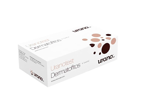
Uranotest Dermatophytes is based on a change in the medium from yellow to res in colour when there is a growth of colonies of the aforementioned fungi. The colour change occurs from the second day after incubation onwards when the dish is incubated at 28º C. At room temperature, the colour change occurs within 12 days at the latest. After this period of the time, any colour change should not be considered as positive.
In contrast to other tube-type dermatophyte tests, Uranotest Dermatophytes is in the shape of a dish which aids the process of seeding the sample and allows the colonies to grow without overlapping each other, thereby making viewing easier. In this way, it aids identificacio and allows an easy test sample of the colonies to be taken using a celophane paper for their re-seeding or observation under a microscope.
- 4 dishes with a DTM culture medium cobered with an aluminium foil.
- 1 bottle with a contrast medium to aid the identification of colonies on observing them under microscope.
- 1 prospectus with instructions for use.
- For beterinary use only.
- For optimum results, adhere strictly to the instructions for use.
- All samples should be handled as if they were potentially infectious and destroyed in accordance with the regulations in force.
- Do not remove the aluminium foil covering each dish until the moment of use.
- Do not reuse.
- Do not use once the expiry date has been passed.
Dermatophytes:
- Affect keratinised tissues: skin, nails and corneal stratum.
- Their diagnosis is very important due to their zoonotic potential.
- Prevalence is greatly increased due to the importing of puppies in a dubious state of health, an increase in adoptions and a rise in the habit of keeping rodents as pets.
Presentation:
- Box of 4 tests.
Includes contrast medium for easy identification and differentiation of the species when observing them under the microscope.


Reviews
There are no reviews yet.